

Summary
Escherichia coli belongs to the Enterobacteriaceae family [1] and is a notable member of normal microbial flora [2]. It is not only the most frequently detected gram-negative rod in septic patients but also a leading cause of community-acquired urinary tract infections [2].
Escherichia coli belongs to the Enterobacteriaceae family [1] and is a notable member of normal microbial flora [2]. It is not only the most frequently detected gram-negative rod in septic patients but also a leading cause of community-acquired urinary tract infections [2]
Staining and Microbiologic Features:
- Gram-negative rod [1]
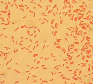
File:E choli Gram.JPG” by Bobjgalindo is licensed under CC BY-SA 4.0.
- E. coli is a lactose-fermenting organism [2].
- It can produce indole as a byproduct of tryptophan metabolism. Therefore, it tests positive for indole [3].
- It exhibits beta-hemolysis when grown on blood agar [3].
- The majority of E. coli strains also test positive for β-glucuronidase. The MUG test uses 4-methylumbelliferyl-β-D-glucuronide (MUG) as a substrate to detect the production of β-glucuronidase [4].
- E. coli is a facultative anaerobe, i.e., it can grow in both the presence and absence of oxygen [5].
- It tests negative for oxidase [5].
- E. coli colonies on EMB agar have a distinctive metallic green sheen [2].
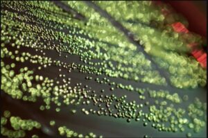
Escherichia coli EMB” by Gene Drendel is licensed under CC BY 4.0.
- E. coli is primarily found in the intestines of humans. So, its detection in water resources suggests fecal contamination of water [2].
- Unlike other E. coli, enterohemorrhagic E. coli does not exhibit sorbitol fermentation [6].
Virulence:
- Non-pathogenic E. coli strains reside in the colon without directly causing gastrointestinal illness. However, they can acquire virulence factors through various mechanisms like conjugation, lysogenic conversion, or transposon-mediated insertion, potentially turning them into pathogenic strains [7].
- E. coli uses fimbriae/pili to latch onto host cells. These structures are helpful in its colonization and infection [3,7].
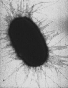
E. coli fimbriae” by (Image: Manu Forero) is licensed under CC BY 2.5.
- K antigen can also aid in bacterial attachment to host cells [8].
- It can use a type III secretion system to release its virulence factors into the target cells [8].
- It produces a siderophore that chelates iron from host proteins, making it available for the bacteria [7].
- Heat-labile Enterotoxin (LT) and Heat-stable Enterotoxin (ST) elevate the levels of cAMP and cGMP, respectively. They promote water and electrolyte loss through enhanced chloride and bicarbonate secretion with decreased sodium and glucose absorption [7].
- Shiga-like toxin disrupts protein synthesis by inactivating the 60S subunit of ribosomes, leading to cell death and tissue damage [7].
- Some other virulence factors include adhesins, flagella, and endotoxin [3].
Transmission:
- Transmission occurs through the fecal-oral route [3] (such as by consumption of contaminated food and water).
- E. coli resides in the colon. However, it can travel up the urethra and cause UTIs [10].
- It can colonize medical equipment such as urinary catheters and central lines and cause nosocomial infections [3].
- At times, bacteria from the oral cavity may be aspirated into the respiratory tract, leading to infection [3]
Diseases and Complications:
- Traveler’s Diarrhea: Heat-labile Enterotoxin (LT) and Heat-stable Enterotoxin (ST) produced by enterotoxigenic E. coli result in severe watery diarrhea [7,11]. “Montezuma’s Revenge” is a colloquial term for traveler’s diarrhea [7].
- Hemorrhagic Colitis: Enterohemorrhagic E. coli produces Shiga-like toxin, leading to hemorrhagic colitis. Affected patients present with abdominal cramps and bloody diarrhea, without fever or the presence of pus in the stool [7].
- Hemolytic Uremic Syndrome: E. coli O157:H7 infection is also associated with hemolytic uremic syndrome. The affected patient may present with a triad of symptoms, including hemolytic anemia, thrombocytopenia, and kidney injury [7]
- As Enteroinvasive E. coli invade and destroy epithelial cells, the body’s immune system mounts an inflammatory response, leading to symptoms like fever, abdominal pain, bloody diarrhea, and pus in stools [7,11].
- Enteroaggregative Escherichia coli is recognized for its capacity to adhere to and aggregate on the intestinal mucosa, resulting in the formation of a distinctive stacked-brick pattern on epithelial cells of the intestine. This strain is implicated in both chronic diarrhea and growth retardation, particularly affecting children. [12]
- It can also cause neonatal meningitis, sepsis, and hospital-acquired pneumonia [10].
Diagnostic Testing:
- Gram staining will show gram-negative rods.
- Culture: E. coli colonies on EMB agar have a distinctive metallic green sheen [2].
E. coli on Eosin-Methylene Blue (EMB) Agar Plate showing growth with green-metallic sheen colonies” by Ajay Kumar Chaurasiya is licensed under CC BY-SA 4.0.
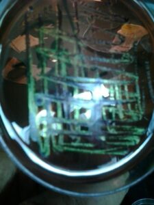
As E. coli can ferment lactose, its colonies have a pink-purple coloration on MacConkey agar [2,11].
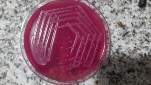
Escherichia coli growth on MacConkey agar” by Ajay Kumar Chaurasiya is licensed under CC BY-SA 4.0.
- Nucleic Acid Amplification Tests (NAATs) [13]
References:
1 First Aid for USMLE step 1, 2021 edition (page no: 145)
2 CMMRS edition 6, 2016-17 (page no: 72)
3 CMMRS edition 6, 2016-17 (page no: 82)
4 Jawetz, Melnick, & Adelberg’s Medical Microbiology Twenty-Seventh Edition (page no: 232, 233)
5 Medical Microbiology by Patrick R. Murray Ph.D., Ken Rosenthal Ph.D., Michael A. Pfaller MD, 8th edition (page no: 253)
6 First Aid for USMLE step 1, 2021 edition (page no: 145)
7 CMMRS edition 6, 2016-17 (page no: 74)
8 Jawetz, Melnick, & Adelberg’s Medical Microbiology Twenty-Seventh Edition (page no: 233)
9 Medical Microbiology by Patrick R. Murray Ph.D., Ken Rosenthal Ph.D., Michael A. Pfaller MD, 8th edition (page no: 255)
10 CMMRS edition 6, 2016-17 (page no: 75)
11 CMMRS edition 6, 2016-17 (page no: 83)
12 Medical Microbiology by Patrick R. Murray Ph.D., Ken Rosenthal Ph.D., Michael A. Pfaller MD, 8th edition (page no: 257)
13 Jawetz, Melnick, & Adelberg’s Medical Microbiology Twenty-Seventh Edition (page no: 237)