

Summary
Some subspecies of Francisella tularensis are the following [1]:
Subspecies tularensis (type A)-most virulent
Subspecies Holarctica (type B)
subsp. mediaasiatica
- Some subspecies of Francisella tularensis are the following [1]:
- Subspecies tularensis (type A)-most virulent
- Subspecies Holarctica (type B)
- subsp. mediaasiatica
Staining and microbiologic features:
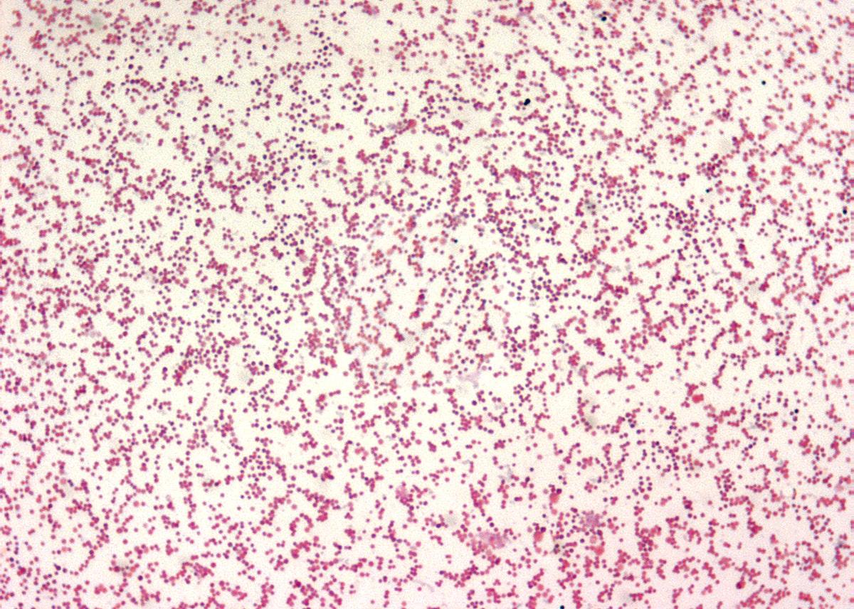
A photomicrograph reveals numerous Gram–negative, Francisella tularensis coccobacilli, the bacterium responsible for causing the disease, tularemia. Original image sourced from US Government department: Public Health Image Library, Centers for Disease Control and Prevention. Under US law this image is copyright free, please credit the government department whenever you can”. by Centers for Disease Control and Prevention is marked with CC0 1.0.
- Facultative intracellular (can survive and replicate in host cells) pathogens that are obligate aerobes [1,3]

File:Macrophage Infected with Francisella tularensis Bacteria (5950310835).jpg by NIAID is licensed under CC BY 2.0.
- Cysteine is required for its growth [2]
- Can be cultured on chocolate agar, modified Thayer-Martin agar, and buffered charcoal yeast extract (BCYE) agar. [4]
- Subspecies tularensis (type A) can ferment glycerol and test positive for citrulline ureidase (unlike subsp. Holarctica/ type B). [4]
Virulence:
- Some mechanisms that aid Francisella in escaping immune response are the following [1]:
- Polysaccharide capsules shield them from phagocytosis and complement-mediated killing.
- Some secreted proteins impair the fusion of phagosomes with lysosomes. Therefore, francisella can escape the lysosomal destruction and replicate in cytosol.
- Compared to other G -ve rods, Francisella endotoxin is less active. [1]
- Highly virulent/infectious and can be a potential weapon for biological warfare.
- Since it is a facultative intracellular organism, B cell-mediated immune response has a limited role in eliminating the organisms. T cell-mediated immune response, IFN-γ, and TNF play an important role in controlling the Francisella infection. [1]
Geographic distribution [1]:
- Less common in the USA
- Type A: Confined to North America.
- Type B: Distributed throughout the northern hemisphere.
Transmission [2]:
- Direct contact while handling infected animal tissues (rabbit/hares)
- Breathing in aerosolized microorganisms
- Arthropods (ticks and deer flies) bite
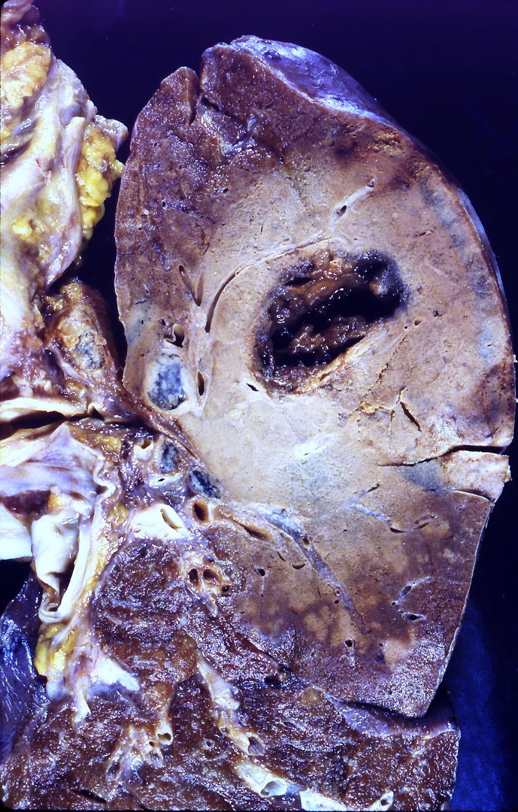
Tularemia (5185046307) by Yale Rosen from USA is licensed under CC BY-SA 2.0.
- Consuming contaminated food or water
Diseases:
- Ulceroglandular tularemia: The patient can develop papules after the initial contact or arthropod bite. These papules are painful and progress to ulcers with a black base. The patient also develops malaise, headache, fever, and painful lymphadenopathy. Symptoms can resemble that of bubonic plague. However, skin ulcers are not a characteristic finding in bubonic plague, and mortality is low in patients with ulceroglandular tularemia. [5,6]
- Pneumonic tularemia: Inhalation of aerosolized microorganisms result in Pneumonic tularemia, and the affected patients have high morbidity and mortality. [5]
Tularemia (5185046307) by Yale Rosen from USA is licensed under CC BY-SA 2.0.
- Exposure of conjunctiva to Francisella tularensis can result in Oculoglandular tularemia. Clinical presentation includes conjunctivitis and preauricular lymphadenopathy. [4,5]
- It can also result in typhoidal tularemia and pharyngeal tularemia. [4]
Diagnostic Testing:
- Small size and faint staining make the organism difficult to visualize on Gram staining. [5]
A 1000X photomicrograph magnification of a direct fluorescent antibody (DFA)–stained specimen, revealing the presence of numerous Francisella tularensis coccobacilli. Original image sourced from US Government department: Public Health Image Library, Centers for Disease Control and Prevention. Under US law this image is copyright free, please credit the government department whenever you can”. by Centers for Disease Control and Prevention is marked with CC0 1.0.
- Culture: Francisella tularensis is a highly infectious pathogen. Therefore, healthcare professionals should follow special measures while collecting and processing the specimen. [5]
- Elevated titers of IgG antibodies against Francisella tularensis [109]
- Skin testing can also detect previous exposure to Francisella tularensis [3]
References:
- Medical Microbiology by Patrick R. Murray Ph.D., Ken Rosenthal Ph.D., Michael A. Pfaller MD, 8th edition (page no: 295)
- CMMRS edition 6, 2016-17 (page no: 108)
- CMMRS edition 6, 2016-17 (page no: 104)
- Jawetz, Melnick, & Adelberg’s Medical Microbiology Twenty-Seventh Edition (page no: 272)
- Medical Microbiology by Patrick R. Murray Ph.D., Ken Rosenthal Ph.D., Michael A. Pfaller MD, 8th edition (page no: 296)
- CMMRS edition 6, 2016-17 (page no: 106)