

Summary
The organism is known for causing plague/black death that resulted in millions of deaths in the past.
Reservoirs for Yersinia pestis include wild rodents, squirrels, prairie dogs, and city rats. [1,2]
The organism is known for causing plague/black death that resulted in millions of deaths in the past.
Staining and microbiologic features:
- Facultative anaerobic bacilli that are gram-negative and non-motile [3]
- Wright, Giemsa, Wayson, or methylene blue staining can be used to visualize Yersinia pestis. [3]
- Safety pin-like/ bipolar appearance on staining [4]

“uu yersinia pestis” by isis325 is licensed under CC BY 2.0.
- Yersinia pestis tests positive for catalase and negative for urease, indole, and oxidase. [4]
- Culture on MacConkey agar yields non-lactose fermenting colonies. [4]
Virulence:
- pla gene and a capsule encoding gene F1 are present on plasmids. [5]
- The protein capsule protects the organism from phagocytosis, and pla protease gene impairs opsonization and chemotaxis of phagocytic cells by disrupting complement components. [5]
- The activity of Plasminogen activating (pla) protease is temperature sensitive. At flea temperature (around 20-28 °C), it shows coagulase activity, and at host body temperature (35-37 °C), it shows fibrinolytic activity and promotes the rapid spread of yersinia. [3]
- Yersiniabactin helps in iron absorption [3]
- Yersinia can directly deliver its virulence factors into host cells using its type III secretion systems. [3]
- Yersinia can evade the phagocytic action of macrophages by dephosphorylating macrophage proteins, destroying macrophage cytoskeleton (especially actin), or promoting its programmed cell death. [5]
- Neutrophils can kill Yersinia pestis. [3]
- Calcium is necessary for its growth at 37 °C. The absence of adequate calcium can alter its metabolic functioning and protein synthesis. [2]
- Some other virulence factors include V and W proteins. [1]
- It can kill other bacteria by producing pesticin [2]
Transmission:
- Yersinia results in zoonotic disease. However, Fleas can serve as a vector, and flea bites can transmit it to humans. [1,2]
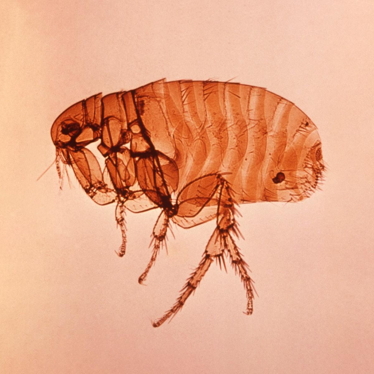
“A close left lateral view of a female Oriental rat flea, Xenopsylla cheopis that transmit the bacterium Yersinia pestis, the causative agent of plague. Original image sourced from US Government department: Public Health Image Library, Centers for Disease Control and Prevention. Under US law this image is copyright free, please credit the government department whenever you can”.” by Centers for Disease Control and Prevention is marked with CC0 1.0.
“Xenopsylla rat flea (plague flea)” by Michael Wunderli is licensed under CC BY 2.0.
- Direct contact while handling infected animals/animal tissues. [2]
- Breathing in aerosolized Yersinia pestis [1,3]
Diseases:
- Bubonic plague: Following an incubation period (that can take up to one week) after the initial exposure, the patient can develop high fever and painful enlargement of lymph nodes in axillary, cervical, and groin regions. Untreated bacteremia can result in hemorrhagic and necrotic lesions throughout the body. Patients may develop disseminated intravascular coagulation and multiorgan failure. [4,6]
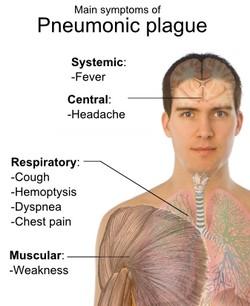
“File:Pneumonic plague.jpg” by Ashfaq is licensed under CC BY-SA 4.0.
- Pneumonic plague: Breathing in aerosolized yersinia pestis results in pneumonic plague. The patient experiences pulmonary symptoms after a short incubation period. This condition is highly contagious and has a very high mortality rate (above 90%).
- Yersinia can also be a potential weapon for biological warfare. [4,6]
Diagnostic testing:
- Gram staining will show Safety pin-like/ bipolar bacilli. [7]
- Culture: Biological safety measures are mandatory while collecting and processing the specimen as the organism is highly contagious.
- Serological testing to detect elevated antibody levels (antibody titer of 1:16 or higher in an unvaccinated patient suggests yersinia pestis infection). [4]
- A rapid diagnostic test for Yersinia pestis detects F1 capsular antigen. [7]
References:
- CMMRS edition 6, 2016-17 (page no: 104)
- CMMRS edition 6, 2016-17 (page no: 108)
- Jawetz, Melnick, & Adelberg’s Medical Microbiology Twenty-Seventh Edition (page no: 275)
- Jawetz, Melnick, & Adelberg’s Medical Microbiology Twenty-Seventh Edition (page no: 276)
- Medical Microbiology by Patrick R. Murray Ph.D., Ken Rosenthal Ph.D., Michael A. Pfaller MD, 8th edition (page no: 261)
- Medical Microbiology by Patrick R. Murray Ph.D., Ken Rosenthal Ph.D., Michael A. Pfaller MD, 8th edition (page no: 262)
- Edition 6, 2016-17 (page no: 109)